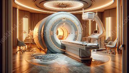
Magnetic Resonance Imaging (MRI) has revolutionized the field of cardiac imaging, providing non-invasive and detailed insights into the heart's structures and functions. In this topic cluster, we will delve into the principles of cardiac imaging with MRI machines, discuss their compatibility with medical devices & equipment, and explore the latest advancements and techniques in the field of magnetic resonance imaging.
Understanding Cardiac Imaging with MRI Machines
Cardiac imaging with MRI machines involves the use of powerful magnets and radio waves to create detailed images of the heart and surrounding blood vessels. Unlike traditional imaging techniques, such as X-rays or CT scans, MRI does not use ionizing radiation, making it a safer option for patients, particularly those requiring repeated imaging.
One of the key advantages of MRI in cardiac imaging is its ability to provide high-resolution, 3D images that capture the intricate structures and dynamic functions of the heart. MRI machines can visualize not only the heart's chambers and valves but also the blood flow, tissue characteristics, and perfusion, allowing for comprehensive assessments of cardiac anatomy and function.
Compatibility with Medical Devices & Equipment
Advancements in MRI technology have led to improved compatibility with various medical devices and equipment. This includes the development of MRI-safe pacemakers, implantable cardioverter defibrillators, and other cardiac devices, allowing patients with these devices to undergo MRI scans safely. Additionally, specialized coils and imaging protocols have been designed to optimize the imaging of patients with implanted devices, ensuring accurate diagnostic results.
Furthermore, MRI machines are compatible with contrast agents that enhance the visualization of cardiac structures and blood flow. These contrast agents, often based on gadolinium, can provide valuable insights into areas of myocardial infarction, inflammation, fibrosis, and perfusion abnormalities, aiding in the diagnosis and management of various cardiac conditions.
Advancements in Magnetic Resonance Imaging
Recent advancements in MRI technology have expanded the capabilities of cardiac imaging. Techniques such as cardiac cine MRI, which captures the motion and function of the heart in real-time, and myocardial tagging, which tracks the deformation of heart muscle, have become integral tools for assessing cardiac function and identifying abnormalities.
Moreover, developments in MRI hardware and software have improved imaging speed, resolution, and image reconstruction techniques, allowing for faster and more precise cardiac scans. The integration of artificial intelligence and machine learning algorithms in MRI analysis has also shown promise in enhancing the interpretation of cardiac MRI images and improving diagnostic accuracy.
The Future of Cardiac Imaging with MRI Machines
Looking ahead, ongoing research and development in cardiac MRI continue to drive innovation in the field. Emerging technologies, such as ultra-high field MRI and novel contrast agents, aim to further enhance the visualization and characterization of cardiac anatomy and pathology.
Additionally, efforts to standardize protocols and establish comprehensive guidelines for cardiac MRI procedures strive to ensure consistent and reliable imaging across different healthcare facilities. This standardization, coupled with the continued evolution of MRI equipment and techniques, is poised to improve the accessibility and quality of cardiac imaging with MRI machines for patients worldwide.


