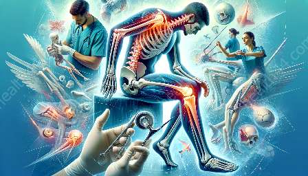
When it comes to diagnosing musculoskeletal injuries and fractures, imaging studies play a critical role in providing accurate, detailed information for orthopedic practitioners. In this article, we'll explore how various imaging techniques are used to diagnose common musculoskeletal injuries, and the real-world applications of these technologies in orthopedics.
The Importance of Imaging Studies
Imaging studies are essential in assessing the extent and nature of musculoskeletal injuries. These studies provide detailed visualizations of bones, joints, ligaments, tendons, and muscles, aiding in the diagnosis and treatment of various orthopedic conditions.
Types of Imaging Studies
Several imaging modalities are commonly used in the diagnosis of musculoskeletal injuries, including X-rays, MRI (Magnetic Resonance Imaging), CT (Computed Tomography) scans, and ultrasound.
X-rays
X-rays are often the first-line imaging tool in the evaluation of musculoskeletal injuries. They provide detailed images of bones, allowing practitioners to identify fractures, dislocations, and other bone-related issues.
MRI
MRI uses strong magnetic fields and radio waves to generate detailed images of soft tissues, ligaments, and tendons. This modality is particularly useful in assessing joint injuries, ligament tears, and soft tissue damage.
CT Scans
CT scans offer cross-sectional images of the body, providing detailed views of bone and soft tissue structures. They are valuable for assessing complex fractures, joint dislocations, and spinal injuries.
Ultrasound
Ultrasound is commonly used to assess soft tissue injuries, such as muscle tears, tendon damage, and joint inflammation. It provides real-time imaging and is often used for guided injections and diagnostic procedures.
Application in Common Musculoskeletal Injuries and Fractures
Imaging studies are crucial in the diagnosis and management of common musculoskeletal injuries and fractures. Let's explore how imaging plays a role in specific conditions:
Fractures
X-rays are the primary imaging tool for assessing fractures, providing detailed views of bone alignment and integrity. CT scans may be used to evaluate complex fractures and assess associated soft tissue injuries.
Sprains and Strains
MRI and ultrasound are valuable in diagnosing ligament and muscle injuries. They provide detailed visualization of soft tissue structures, helping practitioners determine the extent of injury and develop appropriate treatment plans.
Tendon Injuries
MRI and ultrasound are often used to assess tendon injuries, such as tears and inflammation. These modalities aid in accurate diagnosis and guide treatment decisions, including surgical interventions.
Joint Injuries
MRI and CT scans play a critical role in evaluating joint injuries, including detecting cartilage damage, assessing joint stability, and identifying conditions such as osteoarthritis and rheumatoid arthritis.
Real-World Applications in Orthopedics
Imaging studies have revolutionized the field of orthopedics, providing orthopedic surgeons and specialists with the necessary tools to accurately diagnose and treat musculoskeletal injuries. Here are some real-world applications of imaging technology in orthopedics:
Preoperative Planning
Imaging studies allow surgeons to visualize the anatomy of the affected area in detail, aiding in preoperative planning and ensuring precise surgical interventions.
Postoperative Assessment
Following surgical procedures, imaging studies are used to assess the outcomes, monitor healing, and detect any potential complications, guiding the postoperative care of patients.
Guided Injections
Ultrasound imaging is commonly used to guide injections into joints, tendons, and bursae, ensuring accurate delivery of medication and treatment agents to the targeted area.
Monitoring Progress
Imaging studies are essential for monitoring the progress of musculoskeletal injuries and fractures, allowing practitioners to track healing, assess the effectiveness of treatments, and make informed decisions regarding rehabilitation.
Conclusion
Imaging studies play a crucial role in the accurate diagnosis and management of musculoskeletal injuries and fractures in orthopedics. These technologies provide detailed visualizations of internal structures, guiding treatment decisions, and improving patient outcomes. By understanding the real-world applications of imaging technology, orthopedic practitioners can effectively leverage these tools to deliver optimal care to patients.


