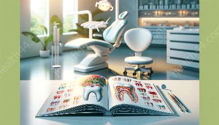
Endodontic therapy, commonly known as root canal treatment, is a dental procedure that involves treating the infected or damaged pulp of the tooth. The pulp chamber, which houses the delicate pulp tissue, plays a significant role in determining the success and challenges of endodontic therapy. The anatomy of the pulp chamber directly impacts the accessibility and intricacies involved in performing root canal procedures.
Anatomy of the Pulp Chamber
The pulp chamber is located at the center of the tooth and extends from the pulp horns to the apical foramen. It comprises the pulp tissue, nerve endings, blood vessels, and connective tissues. The shape and size of the pulp chamber vary from tooth to tooth and can influence the accessibility for endodontic treatment.
Implications for Endodontic Access
The complexity of the pulp chamber's anatomy poses challenges for endodontic access. The location and curvature of the pulp chamber can impact the ability of the endodontist to gain access to the entire pulp system efficiently. Additionally, variations in the number and position of root canals further complicate access and treatment.
Root Canal Treatment and Challenges
During root canal treatment, the endodontist must navigate through the unique anatomy of the pulp chamber to clean, disinfect, and fill the root canals effectively. The accessibility of the pulp chamber greatly determines the success of the treatment and the preservation of the tooth's natural structure.
Consequences of Inadequate Access
If the anatomy of the pulp chamber hinders proper access, it can lead to incomplete cleaning and shaping of the root canals, increasing the risk of residual infection and re-infection. Inadequate access can also compromise the sealing of the root canal, potentially causing treatment failure and the need for retreatment.
Technological Advances and Solutions
Advancements in imaging technology, such as cone-beam computed tomography (CBCT), have facilitated a better understanding of the pulp chamber's anatomy. CBCT scans provide detailed 3D images of the tooth's internal structure, allowing endodontists to analyze the pulp chamber's complexities and plan treatment strategies more effectively.
Furthermore, innovative instrumentation and magnification devices, such as dental operating microscopes, enable enhanced visualization and improved access to the pulp chamber, enhancing the precision and success of endodontic therapy.
Adaptation of Techniques
Endodontic professionals have developed specialized techniques, such as guided access, ultrasonic tips, and rotary instrumentation, to overcome the challenges presented by varying pulp chamber anatomy. These techniques aid in negotiating complex canal systems and ensuring thorough cleaning, shaping, and obturation of the root canals.
Importance of Individualized Approach
Given the diverse nature of pulp chamber anatomy across different teeth and patients, a personalized approach to endodontic therapy is crucial. Endodontists must assess each tooth's unique anatomy and tailor their treatment plan to address specific challenges and ensure optimal outcomes.
Collaborative Care
Collaboration between endodontists and other dental professionals, such as prosthodontists and periodontists, is essential for comprehensive treatment planning and management. Engaging in interdisciplinary discussions allows for a holistic approach to address the complexities of pulp chamber anatomy and its impact on endodontic therapy.
Conclusion
The intricate and diverse anatomy of the pulp chamber significantly influences the accessibility and challenges encountered in endodontic therapy. Understanding the implications of pulp chamber anatomy, embracing technological advancements, adapting specialized techniques, and embracing a personalized and collaborative approach are imperative for successful root canal treatments and the preservation of natural tooth structure.


