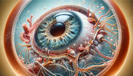
The retina plays a vital role in visual perception, and advancements in retinal imaging technologies have brought about significant implications for the early detection and management of ocular diseases. This topic cluster will delve into the anatomy of the eye, the importance of the retina, and how retinal imaging technologies have revolutionized the diagnosis and treatment of ocular conditions.
Anatomy of the Eye and the Role of Retina
The eye is a complex organ that consists of various structures, each with its own unique function. The retina, located at the back of the eye, is a thin layer of tissue that contains photoreceptor cells responsible for capturing light and transmitting visual information to the brain. It is crucial for creating the images we perceive, making it integral to our vision.
The retina comprises several layers, including the outermost layer known as the retinal pigment epithelium (RPE), which provides vital support to the photoreceptor cells and helps maintain their health. Beneath the RPE lies the sensory retinal layers, which consist of photoreceptor cells (rods and cones), bipolar cells, and ganglion cells. These cells work in concert to convert light into neural signals that are then transmitted through the optic nerve to the brain.
The macula, located at the center of the retina, is responsible for sharp, central vision. Within the macula, there is a small depression called the fovea, which contains a high concentration of cones and is essential for detailed and color vision. The macula and fovea are critical for tasks such as reading, driving, and recognizing faces.
Retinal Imaging Technologies
Retinal imaging technologies encompass a range of sophisticated techniques that allow for non-invasive visualization and analysis of the retina. These technologies have significantly advanced the field of ophthalmology, offering insights into ocular health and aiding in the early detection and monitoring of various eye conditions and diseases.
OCT (Optical Coherence Tomography)
OCT is a non-invasive imaging technique that uses light waves to capture high-resolution, cross-sectional images of the retina. It provides detailed visualization of the retinal layers, allowing for the detection of subtle changes associated with ocular diseases such as macular degeneration, diabetic retinopathy, and glaucoma. OCT has revolutionized the diagnosis and management of retinal conditions by enabling precise measurements of retinal thickness and identifying structural abnormalities.
Fundus Photography
Fundus photography involves capturing digital images of the back of the eye, including the retina, optic disc, and blood vessels, using specialized cameras. These images can aid in the detection and monitoring of retinal pathologies, including diabetic retinopathy, hypertensive retinopathy, and retinal vein occlusions. Fundus photography provides a comprehensive view of the retinal architecture and is often used for documentation and follow-up of ocular diseases.
Fluorescein Angiography
Fluorescein angiography is a diagnostic procedure that involves injecting a fluorescent dye into the bloodstream and capturing images of the retina as the dye circulates through the blood vessels. This technique helps in evaluating the blood flow and identifying abnormal vessel growth or leakage, particularly in conditions such as macular degeneration, diabetic retinopathy, and retinal vascular occlusions. Fluorescein angiography plays a crucial role in determining the extent of retinal pathology and guiding treatment decisions.
Implications for Early Detection and Management of Ocular Diseases
The advent of retinal imaging technologies has transformed the landscape of ocular healthcare, offering numerous implications for the early detection and management of various eye conditions. These technologies enable clinicians to diagnose ocular diseases at earlier stages, allowing for timely intervention and improved outcomes for patients.
Early Detection of Age-Related Macular Degeneration (AMD)
AMD is a leading cause of vision loss in older adults, characterized by degenerative changes in the macula. Retinal imaging technologies such as OCT and fundus photography facilitate the early detection of drusen (yellow deposits beneath the retina) and retinal thinning associated with AMD. Early identification of AMD allows for timely implementation of preventive measures and treatment strategies to slow disease progression and preserve visual function.
Monitoring Diabetic Retinopathy
Diabetic retinopathy is a common complication of diabetes and can lead to vision impairment if left untreated. Retinal imaging techniques, including fundus photography and fluorescein angiography, aid in monitoring the progression of diabetic retinopathy by identifying microaneurysms, hemorrhages, and abnormal blood vessel changes in the retina. Regular retinal imaging plays a pivotal role in early intervention and risk stratification for diabetic retinopathy.
Glaucoma Management and Progression Assessment
Retinal imaging technologies have enhanced the diagnosis and management of glaucoma, a condition characterized by optic nerve damage and peripheral vision loss. OCT enables the measurement of retinal nerve fiber layer thickness, facilitating the early detection of glaucomatous changes. This early detection is instrumental in initiating timely treatment to preserve visual function and halt the progression of the disease.
Paving the Way for Personalized Treatment
Retinal imaging technologies provide clinicians with detailed insights into the structural changes occurring in the retina, allowing for personalized treatment plans tailored to individual patient needs. By identifying specific retinal abnormalities and monitoring their progression, ophthalmologists can customize interventions, such as intravitreal injections, laser therapy, or surgical procedures, to target the precise areas of pathology within the retina.
Conclusion
Retinal imaging technologies have ushered in a new era of ocular healthcare, offering unprecedented opportunities for the early detection and management of various ocular diseases. By leveraging advanced imaging techniques, clinicians can gain valuable insights into the structural and functional changes within the retina, enabling timely intervention and personalized treatment strategies. The implications of these technologies extend beyond diagnosis, shaping the way ophthalmic care is delivered and improving outcomes for patients.


