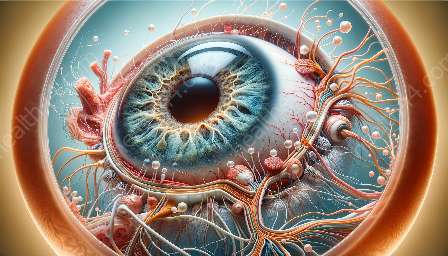
Advancements in imaging technologies play a crucial role in the assessment of the optic disc and the anatomy of the eye. These technologies have evolved over time, enabling healthcare professionals to better diagnose and monitor conditions such as glaucoma, papilledema, and other optic nerve disorders. In this topic cluster, we will explore the interface between imaging technologies and optic disc assessment, delving into the innovative methods that have enhanced our understanding of optic disc anatomy and function.
Understanding the Anatomy of the Optic Disc
The optic disc, also known as the optic nerve head, is the point of exit for ganglion cell axons leaving the eye. It is where the optic nerve enters the eyeball and is devoid of photoreceptors, leading to the blind spot in the visual field. The appearance of the optic disc is essential in diagnosing and managing various ocular and neurological conditions, making accurate assessment crucial for patient care.
Traditional Imaging Methods
Historically, the assessment of the optic disc relied on traditional imaging methods such as direct ophthalmoscopy, fundus photography, and stereoscopic imaging. While these techniques were valuable in their time, they had limitations in terms of resolution, reproducibility, and the ability to capture subtle changes in the optic disc.
Advancements in Imaging Technologies
In recent years, technological advancements have revolutionized the assessment of the optic disc. Imaging modalities such as optical coherence tomography (OCT), confocal scanning laser ophthalmoscopy (CSLO), and scanning laser polarimetry have transformed the way we visualize and analyze the optic disc.
Optical Coherence Tomography (OCT)
OCT is a non-invasive imaging technique that provides high-resolution, cross-sectional images of the optic disc and retina. It allows for the visualization of the retinal nerve fiber layer (RNFL) thickness, optic nerve head morphology, and three-dimensional reconstruction of the optic disc. This level of detail has greatly improved the detection and monitoring of glaucoma and other optic neuropathies, enabling early intervention and better patient outcomes.
Confocal Scanning Laser Ophthalmoscopy (CSLO)
CSLO uses laser light to create high-contrast, high-resolution images of the optic disc. By employing confocal imaging principles, CSLO offers precise visualization of the optic nerve head and surrounding structures. Its ability to detect subtle changes in the optic disc has made it an invaluable tool in the assessment of glaucoma progression and optic disc edema.
Scanning Laser Polarimetry
Scanning laser polarimetry measures the birefringence of the retinal nerve fiber layer to assess the integrity of nerve fibers. This technology quantifies the RNFL thickness and provides data on nerve fiber bundles' structural integrity. It has proven useful in distinguishing between healthy and glaucomatous eyes, aiding in the early diagnosis and management of glaucoma.
Integration with Optic Disc Anatomy
The compatibility of these imaging technologies with optic disc anatomy has significantly enhanced our understanding of the structure and function of the optic nerve head. The ability to visualize the detailed anatomy of the optic disc and measure parameters such as disc size, neuroretinal rim morphology, and RNFL thickness has facilitated the early detection and monitoring of optic disc pathologies.
Future Directions
Advancements in imaging technologies continue to drive progress in optic disc assessment. With ongoing research and development, we anticipate further improvements in resolution, speed, and diagnostic capabilities of imaging modalities. Additionally, the integration of artificial intelligence and machine learning algorithms into image analysis holds promise in automating optic disc assessment, leading to more efficient and accurate diagnoses.
In conclusion, the evolution of imaging technologies for optic disc assessment has revolutionized the way we evaluate and manage various ocular and neurological conditions. The compatibility of these advancements with the anatomy of the optic disc has opened new frontiers in diagnosing and monitoring optic nerve disorders, ultimately improving patient care and visual outcomes.


