Understanding the complex structure and functions of the human eye is crucial for maintaining visual health. From the intricate anatomy of the eye to vision rehabilitation, explore the fascinating world of ocular science.
The Anatomy of the Human Eye
The eye is a remarkable sensory organ that allows humans to perceive the world. The anatomy of the eye can be divided into several components: the external structures, the structure of the eyeball, and the internal structures.
External Structures
The external structures of the eye include the eyelids, eyelashes, and eyebrows, which protect the eye from foreign objects and excessive light. The prominent white part of the eye is called the sclera, while the transparent front part is the cornea, which helps focus light onto the retina.
Within the eye socket, the eye is cushioned by fatty tissue and surrounded by the orbital muscles. The conjunctiva, a thin membrane, covers the sclera and lines the inside of the eyelids.
Structure of the Eyeball
The eyeball is a fluid-filled, spherical structure that houses the delicate components of the eye. The outermost layer is the tough, fibrous sclera, which maintains the shape of the eye and provides attachment points for the eye muscles. The cornea, a clear dome-shaped structure, serves as the window for light to enter the eye.
Beneath the sclera, the middle layer, or uvea, contains the choroid, ciliary body, and iris. The choroid supplies nutrients and oxygen to the retina, while the ciliary body adjusts the lens shape for focusing. The colored part of the eye, the iris, controls the size of the pupil to regulate the amount of light entering the eye.
The innermost layer, the retina, consists of specialized cells called photoreceptors that capture light and convert it into electrical signals for transmission to the brain.
Internal Structures
Inside the eyeball, the anterior chamber is the space between the cornea and the iris, filled with a clear fluid called aqueous humor. The posterior chamber, located behind the iris and in front of the lens, also contains aqueous humor.
The lens, suspended by ligaments, focuses light onto the retina. Behind the lens, the vitreous humor, a clear gel-like substance, maintains the shape of the eye and supports the retina.
Physiology of Vision
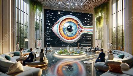
The process of vision begins with the entry of light through the cornea, where it is refracted and passes through the pupil to reach the lens. The lens then focuses the light onto the retina, where photoreceptor cells convert the light into electrical signals.
These signals are transmitted to the brain via the optic nerve, and the brain interprets the signals to create visual images. The photoreceptor cells include rods, which function in low-light conditions, and cones, which enable color vision in bright light.
The visual system integrates the images from each eye to provide depth perception and a comprehensive visual experience. This intricate process involves the coordination of numerous structures and functions within the eye and the brain.
Vision Rehabilitation
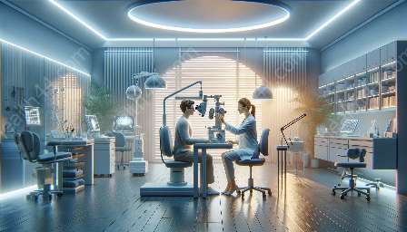
Vision rehabilitation encompasses a range of techniques and interventions aimed at improving visual function in individuals with vision impairments. It addresses the physical, functional, and psychological aspects of vision loss.
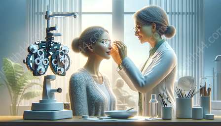
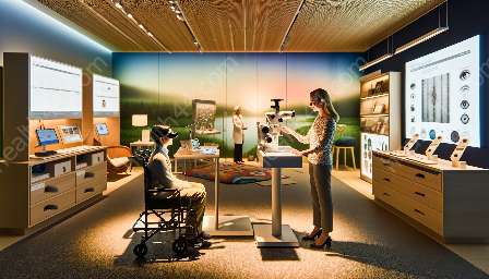
Depending on the nature and severity of the visual impairment, vision rehabilitation may involve optical devices, such as glasses or contact lenses, to correct refractive errors. Additionally, low vision aids, including magnifiers, telescopes, and electronic devices, can enhance visual abilities for daily activities and reading.
Visual training and therapy are also integral components of vision rehabilitation, focusing on improving eye movements, visual processing, and visual integration skills. Occupational therapy and orientation and mobility training help individuals adapt to vision loss and develop strategies for independent living and navigation in the environment.
Furthermore, assistive technology, such as screen readers, voice recognition software, and tactile graphics, supports individuals with visual impairments in accessing information and utilizing digital devices.
Conclusion
The anatomy and physiology of the human eye form the foundation for understanding the mechanisms of vision and the complexities of visual impairments. By appreciating the intricate structures and functions of the eye, as well as the interventions available through vision rehabilitation, individuals can optimize their visual health and well-being.

