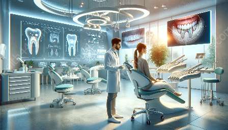
Apexification is a vital aspect of root canal treatment, and advanced imaging modalities play a crucial role in ensuring accurate diagnosis and monitoring of the procedure. This topic cluster aims to explore various imaging modalities such as digital radiography, cone-beam computed tomography (CBCT), and ultrasound, and their significance in apexification and root canal treatment.
The Importance of Imaging Modalities in Apexification
A proper understanding of the internal anatomy of the tooth and periapical structures is essential for successful apexification and root canal treatment. Traditional radiographic techniques have been used for many years, but advancements in imaging technology have led to the development of more sophisticated modalities that offer enhanced diagnostic capabilities.
Digital Radiography
Digital radiography has revolutionized the field of endodontics, providing high-quality images with minimal radiation exposure. It allows for immediate visualization of the tooth and surrounding structures, aiding in the identification of apical pathology and evaluating the progress of apexification. The ability to digitally enhance and manipulate images significantly improves diagnostic accuracy and treatment planning.
Cone-Beam Computed Tomography (CBCT)
Cone-beam computed tomography (CBCT) has gained popularity in endodontics due to its ability to provide three-dimensional images of the tooth and surrounding structures. CBCT offers detailed visualization of the root apex, root canal morphology, and any resorptive defects, allowing endodontists to plan and execute apexification procedures with precision. Furthermore, CBCT is invaluable in identifying the extent of apical pathology and assessing the success of treatment.
Ultrasound Imaging
Ultrasound imaging is a non-invasive modality that can be used to visualize the periapical region and detect changes in the periodontal ligament space. It can aid in the assessment of the healing process following apexification and monitor the development of hard tissue barrier formation. Ultrasound imaging provides real-time feedback, allowing practitioners to make informed decisions during the apexification process.
Integration of Imaging Modalities in Apexification
Combining various imaging modalities offers a comprehensive approach to apexification and root canal treatment. Digital radiography, CBCT, and ultrasound provide complementary information, enabling a thorough assessment of the periapical region and accurate treatment planning. Integrating these modalities enhances the predictability and success rate of apexification procedures, ultimately benefiting the patient's oral health.
Conclusion
In conclusion, the use of advanced imaging modalities in apexification significantly contributes to the success of root canal treatment. Digital radiography, CBCT, and ultrasound play distinct but complementary roles in diagnosing and monitoring apexification, ultimately improving treatment outcomes. As technology continues to advance, the integration of imaging modalities will further enhance the precision and efficacy of apexification procedures, ensuring optimal patient care.


