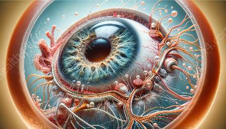
The ciliary muscle is a crucial component of the eye's anatomy, playing a significant role in visual aid design and vision correction. Understanding its implications can lead to the development of more effective visual aids that cater to individual needs. To delve into this topic, it's essential to first explore the anatomy of the ciliary muscle and its relationship to the functioning of the eye.
Anatomy of the Eye
The eye is a marvel of biological engineering, with its intricate structure facilitating the sense of sight. At the core of the eye's anatomy is the ciliary muscle, a key player in the process of accommodation, which enables the eye to focus on objects at various distances. The ciliary muscle is a ring of smooth muscle fibers located within the eye, encircling the lens. It is responsible for altering the shape of the lens to facilitate near or far vision, a process crucial for clear and accurate visual perception.
Function of the Ciliary Muscle
The primary function of the ciliary muscle is to control the shape of the eye's lens, enabling it to refract light and focus on objects at different distances. When the ciliary muscle contracts, it reduces tension on the lens, allowing it to assume a more rounded shape suitable for near vision. Conversely, when the ciliary muscle relaxes, it increases tension on the lens, causing it to flatten and facilitate far vision. This dynamic adjustment of the lens shape, known as accommodation, is essential for clear vision at various distances.
Implications for Visual Aid Design
Understanding the role of the ciliary muscle in accommodation has profound implications for visual aid design. Traditional visual aids, such as eyeglasses and contact lenses, work by correcting refractive errors in the eye by altering the way light enters the eye. However, these solutions may not fully account for the dynamic nature of the ciliary muscle and its impact on the eye's ability to accommodate. As a result, there is a growing interest in developing visual aids that take into consideration the natural mechanisms of accommodation and work in tandem with the ciliary muscle to provide optimal visual correction.
Customized Visual Aids
Advancements in technology have paved the way for customized visual aids that leverage an understanding of ciliary muscle anatomy. For instance, multifocal lenses and adjustable focus eyewear have been designed to accommodate the eye's natural ability to adjust the lens shape for near and distance vision. By incorporating features that mimic the function of the ciliary muscle, these visual aids offer more versatile and personalized vision correction, catering to the individual needs of the wearer.
Presbyopia and Visual Aid Design
Presbyopia, a common age-related condition characterized by the loss of near focusing ability, exemplifies the significance of ciliary muscle anatomy in visual aid design. As the ciliary muscle becomes less flexible with age, the eye's ability to accommodate diminishes, leading to difficulties in seeing objects up close. Understanding the biomechanics of the ciliary muscle has led to the development of progressive lenses and other advanced solutions that address the specific challenges posed by presbyopia, allowing individuals to maintain clear vision at all distances.
Future Innovations
The implications of ciliary muscle anatomy in visual aid design extend beyond current solutions, paving the way for future innovations in vision correction. Researchers and designers are exploring novel approaches, such as accommodating intraocular lenses and dynamic focus eyewear, which aim to integrate seamlessly with the eye's natural accommodation mechanism. These emerging technologies hold the potential to revolutionize vision correction by harnessing the inherent capabilities of the ciliary muscle, thereby providing enhanced visual experiences for individuals of all ages.


