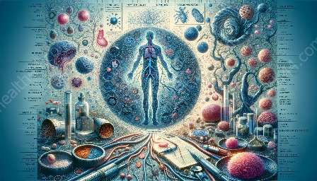
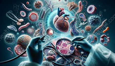
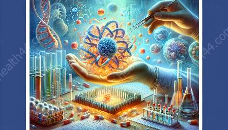
Chemotherapy is a crucial component in the treatment of various malignancies, and its impact on effusions is a key area of interest in cytopathology and pathology. This topic cluster explores the complexities associated with interpreting cytomorphological changes in effusions following chemotherapy, providing insights into the diagnostic and prognostic significance of these alterations.
Understanding Effusions
- Effusion Formation: Effusions, defined as the accumulation of fluid in body cavities, can occur in several anatomical sites, such as the pleural, pericardial, and peritoneal cavities. They are a common manifestation in patients with malignancies.
- Cytological Examination: The evaluation of effusions through cytological examination plays a pivotal role in the diagnosis and management of cancer. This process involves analyzing the cellular components present in the fluid, which can provide valuable information about the underlying pathology.
Impact of Chemotherapy
Chemotherapy exerts profound effects on the cellular composition of effusions, leading to cytomorphological changes that are of great significance in clinical practice. These changes can manifest in various ways, impacting the interpretation of effusion samples and influencing patient management.
Cytomorphological Changes Post Chemotherapy
Post-chemotherapy, effusion samples may exhibit a spectrum of cytomorphological alterations, including changes in cellularity, nuclear features, and cytoplasmic characteristics. These changes can pose challenges in accurately interpreting the malignant potential and treatment responsiveness of the cells within the effusions.
1. Cellularity Changes
Chemotherapy can lead to a reduction in the cell count within effusions. This decrease in cellularity may reflect the cytotoxic effects of the treatment, resulting in a lower yield of abnormal cells for cytological analysis.
2. Nuclear Atypia
The nuclei of cells within effusions may demonstrate varying degrees of atypia following chemotherapy. These alterations can include changes in nuclear size, shape, and chromatin pattern, posing challenges in distinguishing between reactive changes and residual malignancy.
3. Cytoplasmic Alterations
Chemotherapeutic agents can induce changes in the cytoplasmic features of cells present in effusions, such as vacuolization and granularity. These changes may complicate the interpretation of cytological samples, impacting the accurate assessment of cellular characteristics.
Challenges in Interpretation
The cytomorphological changes observed in effusions post chemotherapy present interpretative challenges for cytopathologists and pathologists. Discriminating between treatment-related alterations and residual malignancy requires a comprehensive understanding of the effects of chemotherapy on cellular morphology.
Diagnostic and Prognostic Significance
Accurate interpretation of cytomorphological changes in effusions post chemotherapy is pivotal for informing diagnostic decisions and prognostic assessments. Understanding the implications of these changes can guide subsequent clinical management, including treatment selection and monitoring of therapeutic response.
Future Directions and Research Implications
Advancements in cytopathology and pathology are essential for addressing the evolving landscape of cytomorphological changes in effusions post chemotherapy. Research efforts focused on refining diagnostic criteria and elucidating the underlying biological mechanisms of these changes are imperative for optimizing patient care.
Conclusion
The interpretation of cytomorphological changes in effusions post chemotherapy is a dynamic area within cytopathology and pathology, with direct implications for clinical practice. By comprehensively understanding the complexities associated with these changes, healthcare professionals can enhance their ability to provide accurate diagnostic and prognostic assessments, ultimately improving patient outcomes.


