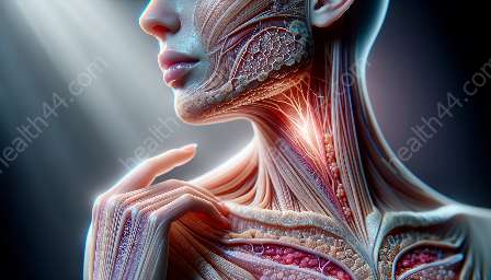
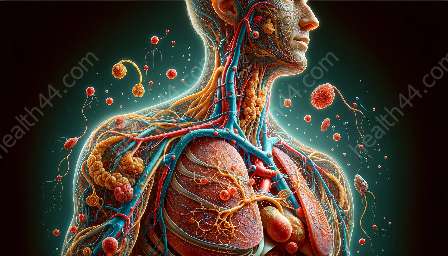
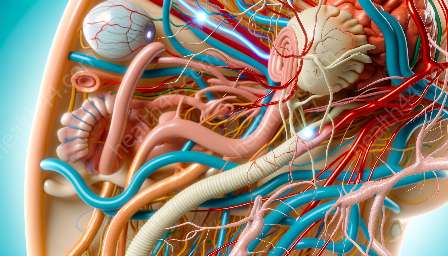
The microscopic anatomy of muscle tissues provides a fascinating insight into the intricate structures and functions of muscle cells. By delving into the microscopic world, we can gain a deeper understanding of the muscular system and its remarkable capabilities.
Types of Muscle Tissues
There are three main types of muscle tissues: skeletal, cardiac, and smooth muscles. Each type has unique characteristics that enable specific functions within the body.
Skeletal Muscle Tissue
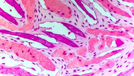
Skeletal muscle tissue is composed of long, cylindrical, multinucleated cells called muscle fibers. These fibers are arranged in parallel bundles and are responsible for voluntary movements and maintaining posture.
Cardiac Muscle Tissue
Cardiac muscle tissue is found in the heart and is responsible for its rhythmic contractions. Unlike skeletal muscle fibers, cardiac muscle cells are branched and interconnected, forming a network that allows coordinated contractions to pump blood throughout the body.
Smooth Muscle Tissue
Smooth muscle tissue is found in the walls of hollow organs such as the digestive tract, blood vessels, and the respiratory system. Its spindle-shaped cells facilitate involuntary movements and help regulate the flow of substances within the body.
Structure of Muscle Cells
At the microscopic level, muscle cells exhibit distinctive features that contribute to their specialized functions. The following are key components of muscle cells:
- Sarcolemma: The plasma membrane of muscle fibers, which surrounds the cell and regulates the passage of materials in and out of the cell.
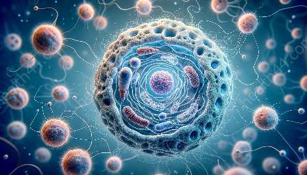
- Sarcoplasm: The cytoplasm of muscle cells, which contains the organelles necessary for muscle contraction, such as myofibrils and mitochondria.
- Myofibrils: Thread-like structures within muscle fibers that contain contractile proteins, including actin and myosin, responsible for muscle contraction.
- Sarcoplasmic Reticulum: A specialized form of endoplasmic reticulum in muscle cells that stores and releases calcium ions, essential for muscle contraction.
- Force Generation: Muscle cells are capable of generating force through the interaction of actin and myosin filaments during contraction, enabling movement and mechanical work.
- Energy Production: Muscle cells possess a high concentration of mitochondria, allowing them to generate energy in the form of adenosine triphosphate (ATP) to support muscle contractions.
- Muscle Hypertrophy: The microscopic analysis of muscle tissues reveals the potential for cells to undergo hypertrophy, increasing in size and strength in response to exercise and resistance training.
Functions of Muscle Cells
Understanding the microscopic anatomy of muscle tissues also sheds light on the vital functions performed by muscle cells. These functions include:
Interplay with the Muscular System
By exploring the microscopic anatomy of muscle tissues, we can connect the intricate details to the broader functions of the muscular system. Muscles work collaboratively to produce movement, maintain posture, generate heat, and support the body's internal organs. The microscopic features of muscle cells directly contribute to these macroscopic functions, highlighting the interconnectedness of the muscular system.
Conclusion
The study of the microscopic anatomy of muscle tissues provides a compelling glimpse into the intricacies of muscle cells and their contributions to the overall functioning of the muscular system. Understanding the types, structures, and functions of muscle tissues enriches our appreciation for the remarkable capabilities of the human body in performing complex movements and maintaining physiological balance.


