Muscle contraction is a complex physiological process that involves the molecular interactions of various proteins and ions, leading to the generation of force and movement in the skeletal muscles of the body.
The Sarcomere: Building Blocks of Muscle Contraction
At the molecular level, the sarcomere is the fundamental unit of skeletal muscle contraction. It is composed of overlapping thick and thin filaments, primarily consisting of myosin and actin proteins.
The myosin filaments contain numerous myosin molecules, each with a head that can bind to actin. Actin filaments are composed of globular actin (G-actin) monomers that polymerize to form the filamentous actin (F-actin). The structural arrangement of myosin and actin within the sarcomere is crucial for the process of muscle contraction.
Sliding Filament Theory
The sliding filament theory explains the mechanism of muscle contraction at the molecular level. According to this theory, during contraction, the myosin heads bind to the actin filaments and pull them towards the center of the sarcomere, leading to a reduction in the length of the sarcomere and the generation of force.
This process involves the release and hydrolysis of adenosine triphosphate (ATP) by the myosin heads, which provides the energy required for the cross-bridge cycle, where the myosin heads repeatedly bind to actin and undergo a conformational change, leading to the sliding of filaments.
Calcium and Muscle Contraction
Calcium ions play a central role in regulating muscle contraction. The release of calcium from the sarcoplasmic reticulum within the muscle fiber is triggered by an action potential, leading to an increase in cytosolic calcium concentration.
Calcium binds to the protein complex troponin, which is associated with the actin filaments, causing a conformational change that exposes the myosin binding sites on actin. This allows the myosin heads to interact with actin, initiating the cross-bridge cycle and leading to muscle contraction.
Neuromuscular Junction and Muscle Contraction
At the neuromuscular junction, acetylcholine released by motor neurons binds to nicotinic acetylcholine receptors on the muscle fiber membrane, leading to depolarization and the generation of an action potential.
This action potential travels along the muscle fiber membrane and into the transverse (T) tubules, leading to the release of calcium from the sarcoplasmic reticulum and the initiation of muscle contraction through the mechanisms described earlier.
Regulation of Muscle Contraction
The process of muscle contraction is tightly regulated by various factors to ensure precise control of muscle movement. These regulatory mechanisms involve the interaction of proteins such as tropomyosin, troponin, and myosin-binding protein C, which modulate the accessibility of myosin binding sites on actin in response to calcium levels and other signals.
Integration with Anatomy and Muscle Function
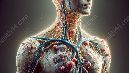
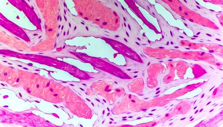
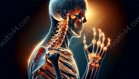
Understanding the molecular basis of muscle contraction is essential for comprehending the anatomical and functional aspects of muscles and movement in the human body. The coordination of sarcomere activity within muscle fibers, the organization of muscle tissue into fascicles, and the attachment of muscles to bones all contribute to the complex interactions that enable efficient movement and physical performance.
By delving into the intricate molecular processes underlying muscle contraction, we gain valuable insights into the dynamic interplay between molecular biology, anatomy, and muscular function, shedding light on the remarkable mechanisms that drive human movement and physical capabilities.

