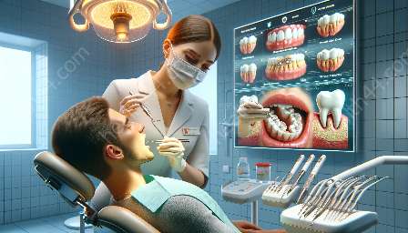
Radiopacity in dental imaging plays a crucial role in the diagnostic process, especially when it comes to assessing the effectiveness of dental fillings and glass ionomer materials. Radiopacity refers to the ability of a substance to block the passage of X-rays, resulting in a brighter appearance on radiographic images. This property is of significant importance in dentistry, as it facilitates the visualization of dental restorations and aids in the detection of caries, fractures, and other dental pathologies.
Understanding Radiopacity
Radiopacity is a fundamental characteristic that allows dental practitioners to distinguish between different anatomical structures and dental materials on radiographs. This property is particularly relevant for materials used in restorative dentistry, such as dental fillings and glass ionomer cements. The ability of these materials to exhibit radiopacity directly impacts their diagnostic value and the overall success of dental treatment.
Significance of Radiopacity in Dental Fillings
Dental fillings, commonly composed of materials like amalgam, composite resins, and other metal alloys, require sufficient radiopacity to be accurately visualized on radiographic images. The radiopacity of these fillings aids in determining their integrity, marginal adaptation, and the presence of secondary caries around the restorations. Without adequate radiopacity, it becomes challenging for dentists to assess the longevity and performance of these restorative materials.
Impact of Radiopacity on Glass Ionomer Materials
Glass ionomer cements are widely used in restorative dentistry due to their unique properties, including adhesion to tooth structure and fluoride release. Their radiopacity is essential for accurate diagnosis, as it enables the differentiation of these materials from tooth structure and surrounding tissues. This distinction is critical in assessing the adaptation and sealing of the glass ionomer restorations, as well as identifying any recurrent caries or debonding issues.
Diagnostic Value of Radiopacity
The diagnostic value of radiopacity in dental imaging extends beyond the visualization of restorative materials. It also aids in the detection of pathologies, such as dental caries and fractures, by providing contrast between the affected structures and surrounding tissues. Additionally, radiopacity plays a key role in treatment planning, as it guides dentists in determining the need for, or success of, additional interventions based on the radiographic findings.
Challenges and Considerations
While radiopacity is essential for accurate diagnosis, certain challenges and considerations need to be addressed. Variations in the radiopacity of dental materials, as well as the thickness and quality of the surrounding tissues, can affect the interpretation of radiographic images. Consequently, dentists must consider these factors and use appropriate imaging techniques to optimize the diagnostic accuracy of radiographs.
Future Implications
Advancements in dental materials and imaging technologies continue to influence the radiopacity and diagnostic value of dental restorations. Ongoing research aims to enhance the radiographic visibility of dental fillings and glass ionomer cements, further improving their diagnostic potential and contributing to enhanced patient care.
- radiopaque substances and their role in dental imaging
- evaluating the radiopacity of dental fillings and glass ionomer materials
- diagnostic significance of radiopacity in dental restorations


