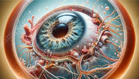
The ciliary body is an essential part of the anatomy of the eye responsible for producing aqueous humor and controlling the shape of the lens. In recent years, significant advancements in imaging techniques have revolutionized our ability to study the ciliary body in detail, leading to a deeper understanding of its structure and function.
Understanding the Anatomy of the Ciliary Body
Before delving into the advancements in imaging techniques, it is crucial to understand the anatomy of the ciliary body. The ciliary body is a ring-shaped tissue structure located behind the iris. It is composed of ciliary processes, ciliary muscle, and the ciliary ring. The ciliary processes are responsible for producing aqueous humor, a clear fluid that nourishes the cornea and the lens, while the ciliary muscle plays a crucial role in controlling the shape of the lens to facilitate near and distance vision.
Advancements in Imaging Techniques
The study of the ciliary body has greatly benefitted from the advancements in imaging technologies. Various imaging modalities have been developed to visualize and study the ciliary body in unprecedented detail.
Optical Coherence Tomography (OCT)
One of the most significant advancements in imaging the ciliary body is the use of optical coherence tomography (OCT). OCT enables non-invasive, high-resolution imaging of the anterior segment of the eye, including the ciliary body. With OCT, researchers and clinicians can visualize the ciliary processes, measure the thickness of the ciliary body, and assess the structure of the ciliary muscle. This technology has provided valuable insights into the dynamics of the ciliary body and its role in various ocular conditions.
Ultrasound Biomicroscopy (UBM)
Ultrasound biomicroscopy is another important imaging technique that has enhanced our understanding of the ciliary body. UBM provides cross-sectional images of the anterior segment of the eye, allowing for the visualization of the ciliary processes, ciliary muscle, and the ciliary body's relationship with other ocular structures. UBM has proven particularly useful in assessing the ciliary body's involvement in conditions such as angle-closure glaucoma and ciliary body tumors.
Anterior Segment Optical Coherence Tomography (AS-OCT)
AS-OCT is a specialized form of OCT that is optimized for imaging the anterior segment of the eye, including the ciliary body. This technique offers high-resolution, real-time imaging of the ciliary body, allowing for quantitative assessment of ciliary body parameters such as thickness and volume. AS-OCT has become a valuable tool in clinical practice for evaluating ciliary body anatomy and pathology.
Implications for Clinical Practice and Research
The advancements in imaging techniques for studying the ciliary body have profound implications for both clinical practice and research. In the clinical setting, these imaging modalities facilitate the diagnosis and monitoring of ciliary body-related conditions such as uveitis, ciliary body cysts, and ciliary body tumors. Additionally, imaging the ciliary body plays a crucial role in the pre-operative assessment of patients undergoing cataract surgery or glaucoma procedures.
From a research perspective, the ability to visualize the ciliary body with unprecedented detail has opened new avenues for understanding the pathophysiology of ocular diseases and developing targeted treatment approaches. Researchers can now investigate the changes in ciliary body morphology and function in various disease states, leading to a deeper understanding of conditions such as angle-closure glaucoma, uveal effusion syndrome, and ciliary body inflammation.
Conclusion
The advancements in imaging techniques have significantly enhanced our ability to study the ciliary body, shedding light on its intricate structure and vital functions within the eye. With the use of cutting-edge imaging modalities such as OCT, UBM, and AS-OCT, researchers and clinicians can now visualize the ciliary body in unprecedented detail, leading to improved diagnosis, treatment, and understanding of ciliary body-related conditions.

