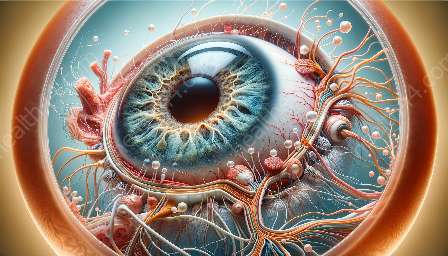
The ciliary body is a complex structure in the eye that plays a crucial role in vision. Variations in ciliary body anatomy can have important clinical implications, affecting ocular health and function. Understanding the intricacies of this anatomy is essential for eye care professionals and individuals interested in ocular health.
Anatomy of the Eye
The eye is a fascinating organ with intricate anatomy designed for the vital function of vision. The ciliary body is a key component of the eye's anatomy, located just behind the iris. It is a ring-shaped structure that connects the iris to the choroid, forming the ciliary muscle and ciliary processes which are involved in the accommodation and production of aqueous humor.
Ciliary Body Structure
The ciliary body consists of three main parts: the ciliary muscle, ciliary processes, and ciliary ring. The ciliary muscle controls the shape of the lens to allow the eye to focus on objects at different distances. The ciliary processes are responsible for producing aqueous humor, a clear fluid that nourishes the cornea and lens. The ciliary ring provides attachment for the ciliary muscle and is essential for maintaining the structural integrity of the ciliary body.
Variations in Ciliary Body Anatomy
Like many anatomical structures in the human body, variations in ciliary body anatomy can occur. These variations may include differences in the size, shape, and position of the ciliary body components, as well as variations in the vascular and nerve supply. Understanding these variations is essential for accurate diagnosis and treatment of ocular conditions.
Impact on Ocular Health
The variations in ciliary body anatomy can have clinical significance in several ways. For example, certain anatomical variations may predispose individuals to develop conditions such as glaucoma or ciliary body cysts. Additionally, variations in ciliary body anatomy may influence the success of certain eye surgeries, such as cataract surgery or glaucoma treatment.
Clinical Evaluation and Management
Eye care professionals utilize various imaging techniques and diagnostic tools to assess the ciliary body anatomy and its variations. This may include optical coherence tomography (OCT), ultrasound biomicroscopy (UBM), and gonioscopy. Understanding the clinical significance of these variations enables ophthalmologists and optometrists to provide personalized and effective management for patients with ocular conditions.
Conclusion
The complexity of ciliary body anatomy and its variations underscores the importance of comprehensive knowledge in the field of ophthalmology and optometry. By understanding the intricacies of the ciliary body, eye care professionals can better diagnose and manage ocular conditions, ultimately contributing to the maintenance of visual health and well-being.


