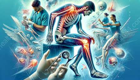
Orthopedic conditions are musculoskeletal conditions that affect the body's bones, joints, muscles, ligaments, tendons, and nerves. Orthopedic imaging techniques play a crucial role in diagnosing and managing these conditions. Among the various imaging modalities, Magnetic Resonance Imaging (MRI) has become an indispensable tool for orthopedic specialists in providing detailed and accurate information about musculoskeletal disorders.
Understanding Orthopedic Imaging Techniques
Orthopedic imaging techniques encompass a wide range of modalities that help in diagnosing and assessing musculoskeletal conditions. These techniques include X-rays, computed tomography (CT), ultrasound, and MRI. Each modality offers distinct advantages and is used based on the specific requirements of the patient and the condition being assessed. However, due to its ability to produce highly detailed images without using ionizing radiation, MRI has gained significant prominence in orthopedic diagnostics.
The Role of MRI in Orthopedics
MRI is a non-invasive imaging technique that uses a powerful magnetic field and radiofrequency pulses to generate detailed images of the body's internal structures. In orthopedics, MRI plays a vital role in evaluating a wide range of musculoskeletal conditions, including bone and soft tissue injuries, degenerative joint diseases, and tumors.
Advantages of MRI in Orthopedics:
- Soft Tissue Visualization: MRI provides exceptional contrast resolution, allowing for clear visualization of soft tissues, including muscles, tendons, ligaments, and cartilage. This capability is particularly valuable in detecting injuries or abnormalities within these structures.
- Multi-Planar Imaging: MRI can produce detailed images in multiple planes, enabling orthopedic specialists to assess the extent and location of abnormalities from various angles, leading to more accurate diagnosis and treatment planning.
- Assessment of Joint Health: MRI is highly effective in evaluating joint structures, such as the articulating surfaces of bones, synovium, and joint capsules. It can identify abnormalities such as tears, inflammation, and degenerative changes, aiding in the diagnosis of conditions like osteoarthritis and rheumatoid arthritis.
- Functional Assessment: Dynamic MRI techniques allow for the assessment of joint and muscle function during movement, providing valuable insights into the biomechanical aspects of orthopedic conditions.
Diagnostic Applications of MRI in Orthopedics
MRI is used in diagnosing a variety of orthopedic conditions, some of which include:
- Rotator Cuff Tears: By offering high-resolution images of the shoulder joint and surrounding tissues, MRI aids in identifying and characterizing rotator cuff tears, guiding appropriate treatment decisions.
- Spinal Disorders: MRI is essential for diagnosing spinal conditions such as herniated discs, spinal stenosis, and spinal tumors, as it provides detailed visualization of the spinal cord, nerve roots, and surrounding structures.
- Joint Injuries: Whether it's a meniscus tear in the knee, ligament sprains, or cartilage injuries, MRI is instrumental in accurately diagnosing and assessing the severity of joint injuries, facilitating timely interventions.
- Tumors and Masses: In the evaluation of musculoskeletal tumors and masses, MRI offers unparalleled tissue characterization and plays a crucial role in determining the nature and extent of the lesion, essential for treatment planning.
- Arthritic Conditions: MRI is widely utilized in assessing arthritis-related changes in joints, aiding in early detection and monitoring disease progression.
How MRI Diagnosis Influences Treatment Decisions
The detailed information obtained from MRI scans significantly influences the treatment approach for orthopedic conditions. These imaging findings help orthopedic surgeons and other healthcare providers tailor treatment plans to address the specific pathology identified. In many cases, MRI results directly impact decisions related to surgery, rehabilitation, or non-invasive interventions.
Case Scenario: Utilization of MRI in Orthopedic Practice
Consider a patient presenting with chronic knee pain and limited range of motion. After a thorough physical examination, the orthopedic specialist orders an MRI of the knee to obtain detailed images of the joint structures and surrounding soft tissues. The MRI reveals a torn meniscus, along with signs of early arthritis. Based on these findings, the orthopedic surgeon can then recommend arthroscopic surgery to address the meniscal tear, along with a customized rehabilitation plan to manage the underlying arthritis.
Advancements in Orthopedic MRI Technology
Ongoing advancements in MRI technology have further enhanced its capabilities in orthopedic imaging. Techniques such as MR arthrography, which involves injecting a contrast agent directly into the joint before imaging, enable improved visualization of intra-articular structures, particularly beneficial for assessing conditions like labral tears in the hip and shoulder. Additionally, advanced sequences and imaging parameters have enabled faster scan times and improved image quality, enhancing the overall efficiency and diagnostic accuracy of orthopedic MRI.
Conclusion
MRI has revolutionized the field of orthopedics by providing precise and detailed insights into musculoskeletal conditions. Its ability to visualize soft tissues, assess joint health, and guide treatment decisions makes it an invaluable tool for orthopedic specialists. As technology continues to evolve, MRI will continue to play a vital role in advancing the diagnosis and management of orthopedic conditions, ultimately improving patient care and outcomes.
By providing comprehensive and actionable information, MRI empowers orthopedic practitioners to deliver personalized and effective care, ultimately enhancing the quality of life for individuals with musculoskeletal ailments.


