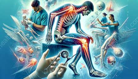
Orthopedic rehabilitation employs various imaging techniques to diagnose and treat musculoskeletal conditions. These imaging methods, including X-rays, MRI, CT scans, and ultrasound, play a vital role in guiding clinicians in the treatment and rehabilitation of orthopedic injuries and conditions.
Orthopedic Imaging Techniques
Orthopedic imaging techniques encompass a range of modalities that are used to visualize the bones, joints, ligaments, tendons, and other musculoskeletal structures. The most common imaging methods used in orthopedics are X-rays, MRI (magnetic resonance imaging), CT (computed tomography) scans, and ultrasound. Each modality offers unique advantages and is employed based on the specific requirements of the patient and the condition being evaluated.
X-rays
X-rays are a widely used imaging technique in orthopedics due to their ability to provide detailed images of bones and joint structures. They are effective in identifying fractures, bone alignment, and degenerative joint diseases. X-rays are quick and convenient, making them a preferred initial imaging method for orthopedic assessments.
MRI
MRI is a powerful imaging tool that uses a magnetic field and radio waves to generate detailed images of soft tissues, muscles, ligaments, and tendons. In orthopedic rehabilitation, MRI is valuable for assessing soft tissue injuries such as ligament tears, tendon damage, and cartilage injuries. It provides high-resolution images without exposing the patient to ionizing radiation, making it particularly useful for certain orthopedic conditions.
CT Scans
CT scans utilize a series of X-ray images taken from different angles to create cross-sectional images of the body's internal structures. In orthopedics, CT scans are used to visualize complex fractures, bone tumors, and joint abnormalities. They offer detailed information about bone density and are instrumental in surgical planning for orthopedic procedures.
Ultrasound
Ultrasound imaging uses high-frequency sound waves to produce real-time images of the musculoskeletal system. It is often employed to evaluate soft tissue injuries, such as tendon and muscle tears, as well as to guide orthopedic procedures such as injections and aspirations. Ultrasound is non-invasive and does not involve ionizing radiation, making it a safe and versatile imaging modality in orthopedic rehabilitation.
Applications of Imaging in Orthopedic Rehabilitation
The applications of imaging in orthopedic rehabilitation are multifaceted and contribute significantly to the diagnosis, treatment, and monitoring of musculoskeletal conditions. Below are key applications of imaging in orthopedic rehabilitation:
- Diagnosis of Orthopedic Injuries: Imaging techniques are crucial for accurately diagnosing orthopedic injuries, including fractures, dislocations, and soft tissue damage. They help clinicians identify the extent and severity of the injury, enabling them to formulate appropriate treatment plans.
- Assessment of Rehabilitation Progress: Following orthopedic interventions, imaging is used to monitor the progress of rehabilitation and healing. By comparing images taken at different time points, clinicians can evaluate the effectiveness of treatment and make adjustments as needed.
- Guiding Orthopedic Interventions: Imaging plays a vital role in guiding orthopedic interventions such as injections, aspirations, and surgical procedures. Accurate visualization of the affected area helps ensure precise placement of injections and surgical instruments, minimizing the risk of complications.
- Planning and Monitoring Postoperative Rehabilitation: For patients undergoing orthopedic surgery, imaging is used to plan the surgical approach and to monitor the healing process postoperatively. It allows clinicians to assess the position of implants, evaluate bone fusion, and detect potential complications.
- Identifying Underlying Pathologies: In cases of chronic musculoskeletal conditions, imaging aids in identifying underlying pathologies such as arthritis, osteoporosis, and soft tissue degeneration. This information is critical for developing comprehensive rehabilitation plans tailored to the individual needs of the patient.
Orthopedics and Imaging Integration
Orthopedics and imaging techniques are intricately integrated, with imaging playing a fundamental role in the practice of orthopedic rehabilitation. The seamless integration of imaging into orthopedic care offers numerous benefits:
- Enhanced Precision in Diagnosis and Treatment: Imaging allows for precise visualization of musculoskeletal structures, enabling accurate diagnosis and targeted treatment planning.
- Minimization of Exploratory Procedures: By providing detailed insights into the nature of orthopedic conditions, imaging reduces the need for invasive exploratory procedures, minimizing patient discomfort and risk.
- Optimization of Rehabilitation Strategies: Imaging findings guide the development of tailored rehabilitation protocols, optimizing the therapeutic approach based on the specific characteristics of the injury or condition.
- Support for Multidisciplinary Collaboration: Imaging results facilitate effective collaboration between orthopedic specialists, radiologists, physical therapists, and other healthcare professionals involved in the rehabilitation process, ensuring comprehensive and cohesive patient care.
Conclusion
The applications of imaging in orthopedic rehabilitation are diverse and indispensable, encompassing crucial roles in diagnosis, treatment planning, and rehabilitation monitoring. Orthopedic imaging techniques empower clinicians to make well-informed decisions and provide personalized care to patients with musculoskeletal injuries and conditions. The integration of imaging into the practice of orthopedics enhances the precision and efficacy of rehabilitation, ultimately contributing to improved patient outcomes and quality of life.


