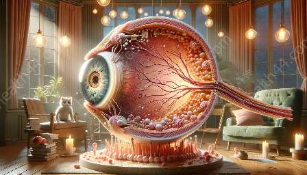
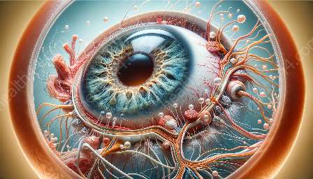
The fovea is a crucial part of the eye's anatomy, playing a significant role in visual function. Any abnormalities in the fovea can have a profound impact on a person's vision and overall quality of life. In this topic cluster, we will explore the foveal abnormalities and their implications on visual function, taking into account the intricate anatomy of the eye.
The Fovea and Its Role in Visual Function
The fovea is a small, central pit within the macula of the retina that is responsible for sharp central vision. Its location and specialized structure enable the eye to focus on fine details and perceive colors with high acuity. The fovea consists mainly of cone cells, which are essential for daylight vision and for recognizing fine details. The surrounding macula, which contains both rods and cones, supports the fovea's function and contributes to overall visual acuity.
Given its critical role in visual function, any abnormalities in the fovea can significantly impact an individual's ability to see clearly, discern details, and perceive colors accurately. These abnormalities can arise from various factors, including genetic predispositions, developmental disorders, and acquired conditions.
Types of Foveal Abnormalities
There are several types of foveal abnormalities that can affect visual function. These include:
- Foveal Hypoplasia: This condition is characterized by the underdevelopment of the fovea, resulting in reduced visual acuity and poor central vision. Foveal hypoplasia can occur in isolation or as part of other syndromes or genetic conditions.
- Foveal Avascular Zone (FAZ) Irregularities: The FAZ is an area within the fovea that lacks blood vessels. Irregularities in the FAZ can lead to decreased visual acuity and distortions in central vision.
- Foveal Cysts: Fluid-filled cysts can develop within the fovea, causing visual distortion, decreased acuity, and difficulty in perceiving fine details.
- Foveal Dystrophy: This progressive condition involves the degeneration of foveal cells, leading to a gradual loss of central vision and color perception.
Each type of foveal abnormality presents its own set of challenges and can significantly impact an individual's visual function and quality of life. Understanding these abnormalities and their implications requires a comprehensive knowledge of the fovea's anatomy and its role in visual processing.
Diagnostic and Management Strategies
Diagnosing foveal abnormalities often involves a thorough ophthalmic examination, including visual acuity tests, optical coherence tomography (OCT) imaging, and fundus photography. These diagnostic tools enable healthcare professionals to assess the structure and health of the fovea and identify any abnormalities that may be present.
Once diagnosed, the management of foveal abnormalities aims to preserve and optimize visual function. This can involve various interventions, such as optical corrections, low-vision aids, and, in some cases, surgical interventions to address specific abnormalities, such as foveal cyst removal or retinal tissue stabilization.
Additionally, ongoing monitoring and regular follow-ups are crucial for individuals with foveal abnormalities to ensure that any changes in visual function are promptly addressed and managed effectively.
Impact on Visual Function and Quality of Life
The impact of foveal abnormalities on visual function extends beyond the physical aspects of vision. Individuals with foveal abnormalities may experience challenges in reading, recognizing faces, navigating their surroundings, and engaging in activities that require precise visual processing. This can have a profound effect on their independence, productivity, and overall well-being.
Furthermore, the psychological and emotional impact of foveal abnormalities should not be overlooked. The frustration and limitations associated with reduced visual acuity and distorted central vision can affect an individual's confidence, self-esteem, and mental health. Therefore, a holistic approach to addressing foveal abnormalities should encompass not only the physical management of visual function but also the psychological and emotional support of the individual.
Conclusion
In conclusion, foveal abnormalities can have far-reaching implications for visual function and the overall quality of life. Understanding the intricate anatomy of the eye, particularly the fovea, is essential for comprehending the impact of these abnormalities and implementing appropriate diagnostic and management strategies.
By recognizing the types of foveal abnormalities and their effects on visual function, healthcare professionals can provide tailored care and support to individuals with these conditions, helping them navigate the challenges associated with reduced central vision and preserving their independence and well-being.


