Visual information is processed and transmitted from the fovea to the visual cortex through intricate neural connections, allowing us to experience sharp central vision. Understanding the anatomy of the eye and the role of the fovea in visual processing is crucial to appreciating the complexities of the visual system.
The Fovea: Center of Visual Acuity
The fovea is a small depression in the retina of the eye, responsible for providing the sharpest and most detailed vision. It is densely packed with cone photoreceptor cells, specialized for color vision and high spatial acuity. Located at the center of the macula, the fovea plays a critical role in focusing light onto the photoreceptor cells to capture fine visual details.
Anatomy of the Eye and Foveal Connections
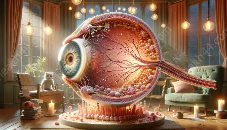
The eye is an intricate organ comprised of various structures that work seamlessly to capture and process visual stimuli. Light enters the eye through the cornea and is refracted by the lens, ultimately reaching the retina at the back of the eye. The fovea, positioned within the macula, receives the focused light, triggering neural signals that are relayed to the visual cortex for further processing.
Foveal Connections to the Visual Cortex
The foveal connections to the visual cortex involve a series of neural pathways that transmit visual information for conscious perception. Neurons from the fovea project to the primary visual cortex, also known as V1 or the striate cortex, via the optic nerve and optic tract. From V1, visual signals are then relayed to higher visual processing areas, allowing for object recognition, motion detection, and other complex visual functions.
Processing Visual Information
Upon reaching the visual cortex, the information from the fovea undergoes intricate processing, including feature extraction, edge detection, and spatial organization. This process enables the brain to construct a detailed representation of the visual scene, supporting our ability to perceive and interpret the world around us.
Visual Pathway and Foveal Function
The visual pathway consists of a series of interconnected structures, including the retina, optic nerve, optic chiasm, optic tract, and various visual processing areas within the brain. The fovea's specialized connection to the visual cortex ensures that fine visual details are accurately represented and analyzed, contributing to our ability to see with exceptional clarity and precision.
Conclusion
Appreciating the foveal connections to the visual cortex and understanding the anatomy of the eye provides valuable insight into the marvels of human vision. The intricate neural pathways and processing mechanisms involved in foveal vision highlight the remarkable capabilities of our visual system, emphasizing the importance of preserving and caring for this vital sensory organ.

