The fovea is a critical structure in the human eye responsible for high-acuity vision, and understanding its morphology is vital in the field of ophthalmology and vision science. Additionally, image processing plays a crucial role in enhancing visual information, and when combined with knowledge of foveal morphology, it can lead to significant advancements in various fields, including medicine, technology, and artificial intelligence.
Foveal Morphology and Anatomy of the Eye
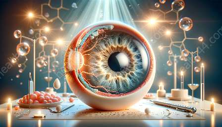
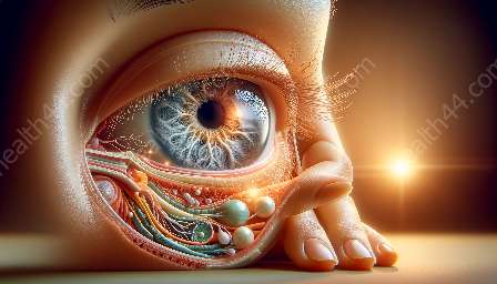
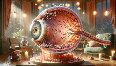
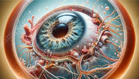
The fovea is a small, central pit in the macula of the retina that is responsible for sharp and detailed vision. It is located at the center of the macula, a specialized part of the retina that provides the clearest vision and the highest visual acuity. The anatomy of the eye, particularly the structure of the fovea, is essential to understand the relationship between foveal morphology and image processing.
The fovea centralis, commonly referred to as the fovea, consists of densely packed cones, the photoreceptor cells responsible for color vision and visual acuity. This high concentration of cones in the fovea allows for the perception of fine details and is crucial for tasks such as reading, driving, and recognizing faces. The fovea's unique structure and high density of cones make it a key area for researchers and medical professionals aiming to understand and treat various ocular conditions.
Importance of Foveal Morphology
Foveal morphology is of paramount importance in the study of vision, as the structure of the fovea directly impacts an individual's ability to perceive fine details and achieve high-resolution vision. The high density of cones in the fovea results in a higher resolution of visual information, which contributes to the human ability to focus on and distinguish small or subtle details within their field of view.
Understanding foveal morphology is particularly crucial in the diagnosis and management of retinal diseases and conditions that affect central vision. Diseases such as age-related macular degeneration (AMD), diabetic retinopathy, and macular edema can significantly impact the foveal region, leading to a decline in visual acuity and overall vision quality. By comprehensively studying foveal morphology, researchers and clinicians can develop targeted treatments and interventions to preserve and enhance foveal function.
Role of Image Processing in Enhancing Visual Information
Image processing is a multidisciplinary field that focuses on analyzing and manipulating visual information to improve its quality, extract useful data, or make it more suitable for specific applications. In the context of foveal morphology and the anatomy of the eye, image processing techniques can be employed to enhance visual information received by the fovea and improve overall visual perception.
One of the primary goals of image processing in relation to foveal morphology is to optimize the presentation of visual stimuli to the fovea, thus maximizing the information processed by the high-density cone cells. By carefully adjusting the contrast, brightness, and spatial characteristics of visual stimuli, image processing techniques can help individuals with compromised foveal function receive and interpret visual information more effectively.
Integration of Foveal Morphology and Image Processing
The integration of foveal morphology and image processing holds significant promise in various fields. In the domain of medicine, understanding the intricate structure of the fovea alongside advanced image processing methodologies can aid in the earlier diagnosis and monitoring of retinal diseases. By analyzing and enhancing images of the fovea, medical professionals can detect subtle changes in foveal morphology, leading to timely interventions and personalized treatment plans.
Furthermore, the integration of foveal morphology and image processing has implications for innovative technological applications. In the development of virtual reality (VR) and augmented reality (AR) systems, an in-depth understanding of foveal morphology can influence the design of visual interfaces to maximize user engagement and perceptual immersion. Image processing algorithms can be utilized to adapt virtual visual content, such as text and graphics, in a manner that aligns with the fovea's sensitivity to detail and color.
Advancements in Artificial Intelligence
The intersection of foveal morphology and image processing also extends to the field of artificial intelligence (AI). By emulating the mechanisms of the human fovea and integrating advanced image processing techniques, AI systems can be designed to process visual information with improved efficiency and precision. This is especially relevant in tasks such as object recognition, where mimicking the foveal function through image processing algorithms can lead to more accurate and rapid identification of objects within complex visual scenes.
The symbiotic relationship between foveal morphology and image processing presents a fertile ground for interdisciplinary collaboration, where expertise in ophthalmology, neuroscience, computer science, and engineering converges to drive scientific breakthroughs and technological innovations.
Future Prospects and Conclusion
The exploration of foveal morphology and its synergy with image processing opens up avenues for further research and practical applications. In the coming years, advancements in imaging technologies, computational algorithms, and medical interventions will continue to leverage the profound insights gained from understanding foveal anatomy and implementing image processing techniques.
In conclusion, foveal morphology and image processing intertwine to enrich our understanding of vision, enable medical advancements, and inspire the development of cutting-edge technologies. Embracing these interconnected domains has the potential to empower individuals with visual impairments, enhance human-computer interaction, and advance the frontiers of artificial intelligence. By recognizing the intricate relationship between the fovea, image processing, and the anatomy of the eye, we can harness the full potential of these disciplines to shape a clearer and more visually captivating future.

